Home » Archives for THS Hospital » Page 12
Home » Archives for THS Hospital » Page 12
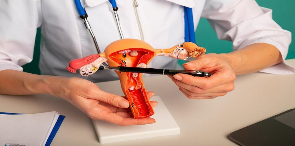
HPV Vaccination
V
accination has proven to be a successful measure in the prevention of multiple infectious diseases and its massive application has achieved the disappearance of many of them. An infectious agent is an indispensable part of the genesis of cervical cancer and the idea of looking for a vaccine against that agent came a long time ago.
Currently, the vaccine is a reality: the infectious agent is human papillomavirus (HPV) of a variety of high oncogenic risks. Human papillomavirus vaccines are a new and effective resource in the strategies of primary prevention of cervical cancer.
Effectiveness of vaccines against cervical cancer precursor lesions:
The preventive effect of the available human papillomavirus vaccines is proven for precursor lesions of cervical cancer.
There are three stages of cervical intraepithelial neoplasia (CIN): CIN 1, CIN 2, and CIN 3. These stages are determined according to the severity of the changes that appear in the cells: slight, moderate, or high.
The tetravalent vaccine protects against CIN 2 produced by human papillomaviruses 16 and 18, in 97 to 100% of women. For cross-protection, it also protects against the same injuries caused by other types of high-risk human papillomaviruses: type 31 (46.2% of protection), type 33 (28.7%), type 45, (7.8%) and type 52 (18.4 % of protection).
The bivalent vaccine protects against CIN 2 produced by human papillomavirus 16 and 18 in around 92.9 and 95.7% of women. For cross-protection, it also protects against injuries caused by types 31 (36.1% of protection), 33 (36.5%), 45 (59.9%) and 52 (31.6%).
Despite their high rates of effectiveness, vaccinated women may develop cervical cancer due to vaccine failure (which is extremely rare) or because it is caused by high-risk human papillomavirus against which it was not offered protection. The need for detection, even among vaccinated women, it’s essential.
Other benefits of human papillomavirus vaccination
The human papillomavirus vaccination offers, at the same time to vaccinated men and women, protection against other types of cancer.
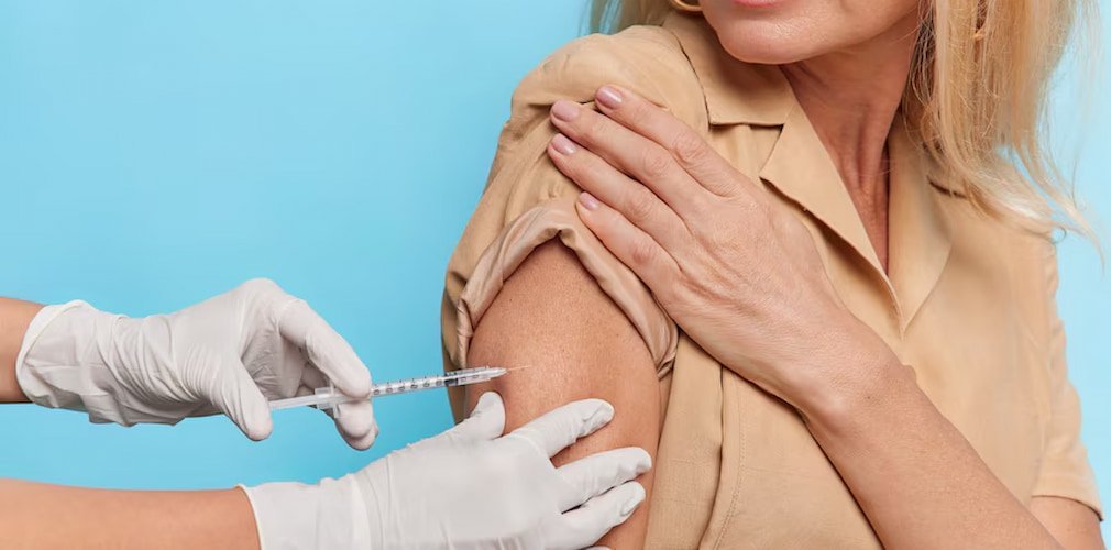
Patients can get a vaccine against HPV at Harley Street Hospital. Book an appointment with us to help prevent this disease.
Frequently Asked Questions
Yes, the HPV vaccine is approved for individuals up to age 45 in some countries, but effectiveness may decrease with age. It is best to consult with a healthcare provider to determine if it is appropriate for you.
The HPV vaccine protects against certain strains of the human papillomavirus (HPV) that can cause cervical cancer, genital warts, and other HPV-related cancers.
The HPV vaccine is typically recommended for both males and females between the ages of 9 and 26, with the possibility of vaccination up to age 45 in some cases.
The HPV vaccine is ideally administered before the onset of sexual activity, typically between ages 11 and 12, but it can be given up to age 26 for those who have not been vaccinated previously or completed the series.