Posterior Cervical Spine Surgery


Posterior Cervical Spine Surgery

C

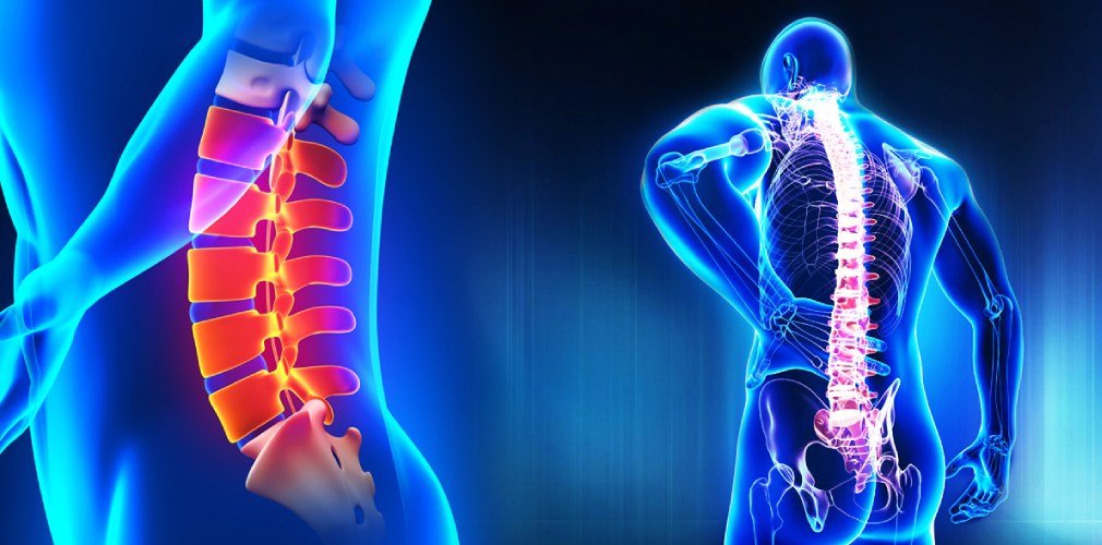
Cervical Spine Surgery may be indicated for patients with trauma, degeneration, persistent neck pain, and unstable conditions. Surgery to the cervical spine should be undertaken by specialists with experience.

Specialists in Cervical Spine Surgery

Many neck problems are due to degenerative changes that occur in the intervertebral discs of the cervical spine and the joints between each vertebra. Other problems are the result of injury to parts of the spine or complications of earlier surgeries. The vast majority of patients who have neck problems will not require any type of operation. However, if the non-operative treatments fail to control your pain or problems, your spine surgeon may suggest a posterior cervical fusion to treat your neck problem.

The purpose of this information is to help you understand:

The anatomy of the cervical spine

  • The types of problems a posterior cervical fusion is used for
  • The rationale for performing a posterior cervical fusion
  • What you can expect from this procedure
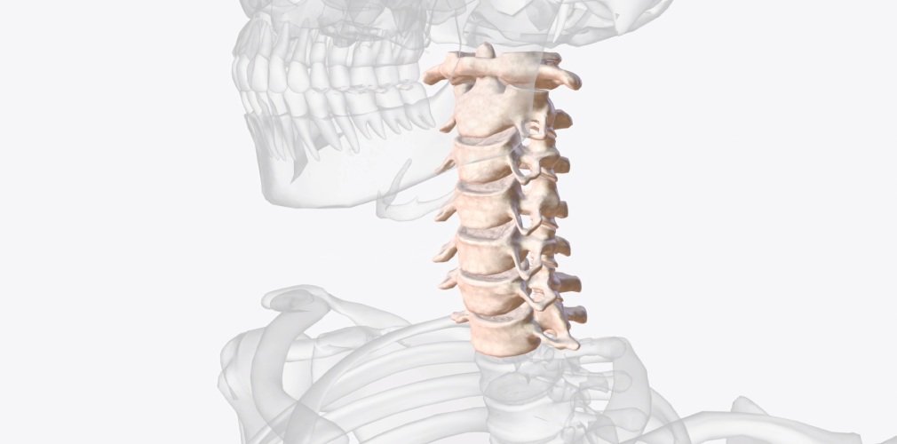
Anatomy

In order to understand your symptoms and treatment options, you should start with some understanding of the general anatomy of your neck. This includes becoming familiar with the various parts that make up the neck and how these parts work together.

Surgery is not necessary in every case. No one type of surgery works for every neck pain problem. Numerous surgical procedures have been designed to treat each type of neck pain.

A posterior cervical fusion may be recommended for several reasons:

  • To stop the motion between two or more vertebrae – or spinal segments
  • To straighten the cervical spine and stop the progression of a spinal deformity
  • To stabilize the spine after a fracture or dislocation of the cervical spine

The goal of a posterior spinal fusion is to allow two or more vertebrae to grow together or fuse into one solid bone. If the operation is being done because you are suffering from mechanical neck pain, the fusion can stop the excess motion between the vertebrae caused by segmental instability. This can reduce your pain.

If your spine is unstable, due to a fracture or a dislocation of the vertebrae, the fusion stabilizes the spine. This is particularly important when you have not injured your spinal cord. Stabilizing the spine surgically can protect you from spinal cord injury during the healing process. Even in cases when there has been severe damage to the spinal cord leading to paralysis, a spinal fusion may be recommended, so that you can get out of bed and into a wheelchair faster. This allows early rehabilitation because the spine has been stabilized by the internal fixation.

A posterior cervical fusion may also be suggested to straighten the spine, or control a deformity of the cervical spine such as a cervical kyphosis. The cervical spine normally has a “C” shaped curve with the opening towards the back. A “kyphotic” curve is exactly the opposite – the opening of the “C” points forward. This deformity occurs when the cervical spine is unstable and begins to bend forward.

The Operation

Posterior Cervical Fusion

The bone graft is usually taken from the pelvis at the time of surgery, but some surgeons prefer to use bone graft obtained from a bone bank. Bone graft from a bone bank is taken from organ donors and stored under sterile conditions until needed for operations such as spinal fusion. The bone goes through a rigorous testing procedure, similar to a blood transfusion. This is in order to reduce the risk of passing on diseases, such as AIDS or hepatitis, to the recipient.

There are two basic types of cervical spinal fusion:

Anterior Interbody Fusion

This type of fusion is much more common in the neck. This type of fusion is described above. In the interbody fusion, a bone graft is placed between two vertebrae and replaces the removed disc. During the healing process, the vertebrae grow together, creating a solid piece of bone out of the two vertebrae.

Posterior Fusion

In the posterior fusion, the bone graft is placed on the backside of the vertebrae. During the healing process, the vertebrae grow together, creating a solid piece of bone out of the two vertebrae. This type of fusion is used in the cervical spine for fractures and dislocations of the cervical spine, and to correct deformities in the neck such as cervical kyphosis.

Instrumented Posterior Cervical Fusion

When doing an anterior cervical fusion, the bone graft may simply be wedged in between the vertebra. It is held there simply because it is wedged in tight. In the case of a posterior cervical spine fusion, there is not anywhere to wedge bone graft material, so the bone graft is simply laid on top of the lamina of the vertebrae.

In recent years, there has been an increase in the use of metal plates, screws, and rods to try to increase the success of helping the spine to fuse. Many different types of metal implants are used; all try to hold the vertebrae in position while the fusion heals. Bone heals best when it is held still, without motion between the pieces trying to heal together. The healing of a fusion is no different than healing a fractured bone, such as a broken arm. However, the neck is a difficult part of the body to hold still.

In the past, casts and braces were used in an attempt to reduce the motion in the neck and to increase the success rates of a spinal fusion. In most cases, these braces and casts were simply too cumbersome to wear for three months, and did a poor job of actually holding the neck still enough to allow the fusion to heal.

In the posterior cervical spine fusion, it is common to use stainless steel wire to hold the bones together as they heal. The wires are wrapped around the spinous processes, or under the lamina and twisted together. This creates a fairly strong form of internal fixation that holds the bones in place while they heal.

By using wires, the vertebra can be held rigidly in place while the fusion heals. Braces and casts are not needed.

Complications

With any surgery, there is a risk of complications. When surgery is done near the spine and spinal cord these complications (if they occur) can be very serious. Complications could involve subsequent pain and impairment and the need for additional surgery. You should discuss the complications associated with surgery with your doctor before surgery. The list of complications provided here is not intended to be a complete list of complications and is not a substitute for discussing the risks of surgery with your doctor. Only your doctor can evaluate your condition and inform you of the risks of any medical treatment he or she may recommend.

Frequently Asked Questions

Posterior cervical spine surgery is a surgical procedure performed on the back of the neck to address conditions affecting the cervical spine, such as herniated discs, spinal stenosis, or degenerative disc disease. It involves accessing the spine through the back of the neck to relieve pressure on the spinal cord or nerves and stabilise the affected area.

Common reasons for posterior cervical spine surgery include severe neck pain, weakness, or numbness in the arms, difficulty walking, or loss of bladder or bowel control due to compression of the spinal cord or nerves. Surgery may be recommended when conservative treatments such as medication, physical therapy, or injections fail to provide adequate relief.

Recovery from posterior cervical spine surgery varies depending on the specific procedure performed and individual factors. It typically involves a hospital stay of a few days, followed by a period of restricted activity and physical therapy to regain strength and mobility. Pain management and adherence to post-operative instructions are essential for optimising recovery and achieving the best possible outcome.

Doctors Specialising in Spine Care

Day Surgery Lumbar Microdiscectomy

L

umbar microdiscectomy basically consists of a small surgery to remove the herniated part of the lumbar disc. This injury to the disc causes irritation and/or inflammation in the nerve root since the disc puts pressure on it.

Normally, when the herniated disc produces intense pain, radically affecting our daily life, the intervention is chosen. Microdiscectomy is normally carried out for a lumbar disc herniation, it is highly effective against the leg pain that it causes.

What should I consider before a lumbar microdiscectomy?
Before talking to you about the surgical procedure, it is important to carry out a series of guidelines in our everyday life. Therefore, we are going to leave you with a series of recommendations to deal with lumbar microdiscectomy and its subsequent recovery:

  • Avoid smoking before surgery.
  • Be careful with certain medications. Before surgery, it is essential to inform if you are taking medication.
  • Leading a healthy lifestyle will help the subsequent recovery.
  • During the first days after surgery, it is recommended to make the minimum effort.
  • You should consider asking someone for help in getting you from the hospital to your home, as well as for daily tasks.

The surgical procedure
Having given these tips, it is time to clear up doubts about the surgical procedure:

  • The surgeon will make a small incision, that is, a cut in the midline of the lower back.
  • Next, some of the muscles are separated in order to expose the back and have the correct access to the spine.
  • The surgeon will make a small hole called a laminectomy in your spine.
  • Using the microscope, the surgeon will identify the nerves and disc.
  • Once all the above procedure is done, it is time to remove the damaged part of the disc.
  • Once the pressure on the nerve has been removed, the surgeon will suture the wound.

The results
We must consider that a herniated disc that causes almost excruciating leg pain can take weeks to improve. However, patients who undergo this type of intervention allege that they feel relief in their legs shortly after the intervention.

Another fact to keep in mind is that between 85-90% of the people intervened allege that the intervention has been worthwhile.

Advantages of lumbar microdiscectomy

  • We will mention the following:
  • It decreases leg pain caused by a herniated disc, even eliminating it.
  • The incision made is very small, making it minimally invasive. The surgical incision is at most 3 cms.
  • The healthy material of the disc does not suffer, that is, it is conserved and continues to function normally.
  • Adjacent discs are not damaged in any way.
  • On the day of surgery, you will be able to walk and get up.
  • A quick return to activities of daily and work life.

Frequently Asked Questions

Yes, microdiscectomy is often performed as an outpatient procedure, allowing patients to return home on the same day as the surgery in many cases.

Some back surgeries, such as minimally invasive procedures or certain spinal decompression surgeries, can be performed as day surgeries, allowing patients to return home the same day, while more complex procedures may require a hospital stay for observation and recovery.

A microdiscectomy typically takes around 1 to 2 hours, although the exact duration may vary depending on factors such as the complexity of the procedure and individual patient factors.

Doctors Specialising in Spine Care

Day Surgery Cervical Disc Replacement

G

enerally, the discomfort caused by degenerative diseases of the spine is treated with anti-inflammatory drugs, physical therapy and/or infiltrations. However, sometimes the only way to relieve pain and regain mobility is through surgery (cervical disc replacement).

There is a novel surgical technique to treat the most complex cases of cervical disc herniation, a degenerative disease that affects the cervical discs, which are located between the vertebrae of the spine and function as shock absorbers.

The procedure
This sophisticated procedure, called cervical arthroplasty, involves implanting a prosthesis, which is a flexible, synthetic device designed to replace the affected disc. This method allows faster recovery since it reduces the postoperative time from seven days to one. Also, patients do not need to wear a cervical collar after the procedure.

This new surgical modality obeys the current trend of placing prostheses to restore movement of the diseased joint, a technique widely used for example in cases of osteoarthritis of hip or knee.

Main advantages
In traditional surgeries, the affected disc is replaced by bone and the corresponding vertebrae are fixed using plates, in order to join that segment of the spine.

Unfortunately, these vertebral fusions have some disadvantages. Firstly, since the bone’s ability to heal is variable, it is possible for fusion to fail, which can lead to persistent neck pain. Furthermore, this type of joints can generate stiffness in the operated sector, which causes an increase in the mechanical requirements of the adjacent intervertebral discs.

The problem is that this transmitted overstrain can cause accelerated wear of the affected area, which sometimes results in the need for new surgeries to repair the area.

On the other hand, with the new technique, the prosthesis that is placed is not fixed to the upper and lower vertebrae but fits perfectly between the vertebral bodies.

Frequently Asked Questions

Cervical disc replacement surgery typically requires a short hospital stay of one to two days rather than being performed as a same-day surgery.

Patients may typically begin walking shortly after cervical disc replacement surgery, although specific post-operative instructions and restrictions will vary depending on the individual’s condition and the surgeon’s recommendations.

After cervical disc replacement surgery, it’s important to avoid heavy lifting, bending, or twisting motions that could strain the neck, and to follow post-operative instructions provided by the surgeon to promote proper healing.

Doctors Specialising in Spine Care

Glaucoma

Glaucoma

Glaucoma is the main cause of irreversible blindness in the world.
It is not unknown that glaucoma (acute attack of glaucoma) is a pathology of frequent consultation in the emergency services, because of its sudden onset and because of the acute and intense pain it produces.
The diagnosis must be immediate and accurate, as well as the proper handling, to avoid irreversible sequelae.

Risk factors
The risk factors related to this pathology are:

  • Age: At the sixth decade and with the passage of time the lens increases the anteroposterior diameter, so this reduces progressively the dimensions of the anterior chamber facilitating angular closure.
  • Female sex: because of their lower size and, consequently, smaller eyeball sizes.
  • Family history: involved brothers and fathers.
  • Asian ancestry.

Clinical presentation
The classic clinical presentation is a patient who presents suddenly and acutely, diminution of the visual acuity generally unilateral (only on one eye), with a vision of haloes of colors around the lights; halos that are characterized by being yellow and orange around the lights.
In addition, very intense pain in the affected eye with irradiation to the head region on the same side, accompanied by symptoms such as nausea, vomiting, dizziness, abdominal cramps and palpitations.
Symptoms usually develop in the early morning or during shows that take place with low lighting (cinema, television or restaurants), because of the pupil dilatation generated by the low illumination, which replicates the iris on the angle blocking it.
 
Treatment
The treatment is aimed at sharply reducing intraocular pressure with different drugs.

Frequently Asked Questions

Glaucoma is a group of eye conditions that damage the optic nerve, typically due to increased pressure within the eye, leading to vision loss or blindness if left untreated.

Glaucoma is diagnosed through a comprehensive eye exam, which may include measurement of intraocular pressure, examination of the optic nerve, visual field testing, and evaluation of the drainage angle in the eye.

Treatment for glaucoma aims to lower intraocular pressure to prevent further damage to the optic nerve. This may involve eye drops, oral medications, laser therapy, or surgical procedures such as trabeculectomy or drainage implants. Regular monitoring and adherence to treatment are essential to manage glaucoma effectively.

Diabetic Retinopathy

Diabetic Retinopathy

D

iabetic retinopathy is a condition of the retina caused by Diabetes Mellitus. It appears in Type 1 Diabetes Mellitus in 98% of patients at 20 years of disease and in Type 2 Diabetes Mellitus in 60% at 20 years of diabetes.

The first risk factor for the appearance of this ocular problem that can lead to blindness is to suffer from Diabetes Mellitus in a prolonged way over time. The second risk factor is poor metabolic control, very important for the development and progression of this pathology. The worse control the more severe the diabetic retinopathy will be.

The patient notices loss of vision generally when this pathology has already advanced so it is mandatory and important, the diabetic fundus eye is checked periodically and before the loss of vision begins.

Therefore, the patient may have diabetic retinopathy and not be aware of it for not presenting symptoms. As the progression of the problem occurs, patients may perceive any of the following symptoms:

  • Vision loss
  • Blurry or fluctuating vision (goes from clear to blurred).
  • Bad night vision
  • Difficulty perceiving colors.
  • Stains in the visual field or dark areas (main symptom of retinal detachment).

When the diabetic retinopathy is suspected, the ophthalmologist will perform a study of the fundus with fluorescein angiography, which will allow him to know the characteristics of the alterations of the blood vessels of the retina, and with OCT (Optical Coherence Tomography), which offers information of size, location, volume of exudation (waterlogging) and affected layers of the retina. This test is also very important to study the possible edema of the macula (the macula is the central part of the retina).

The patient must have strict control of glycemia (glycosylated hemoglobin less than 7%).

In the case of patients diagnosed with type 2 diabetes, it is necessary to perform an eye fundus study as soon as possible. In those who suffer from type 1 diabetes, the test should be performed 5 years after diagnosis. Subsequent revisions should be carried out annually if the fundus is normal or if there is mild diabetic retinopathy. If the diabetic retinopathy is moderate, the revisions will be carried out every six months and every four months if a risk of the proliferative form is suspected.

Treatment

The basic treatment of diabetic retinopathy is laser photocoagulation in one or several sessions after checking the state of the blood vessels through a fluorescein angiography. This photocoagulation with a laser can be associated with intravitreal injections of anti-angiogenic medication in some forms with neovessels (newly formed blood vessels). To treat macular edema (build-up of fluid in the macula), intravitreal injections of anti-angiogenic medication will be applied with or without the laser. Corticosteroids can also be used as a treatment inside the eyeball. The possible complications will be treated with surgery (vitrectomy that consists of the extraction of the vitreous humor through different procedures).

Frequently Asked Questions

Diabetic retinopathy is largely preventable through tight control of blood sugar levels, regular eye exams, blood pressure management, and healthy lifestyle choices like maintaining a balanced diet and avoiding smoking. Early detection and timely treatment are key in preventing vision loss.

The four stages of diabetic retinopathy include mild nonproliferative retinopathy, moderate nonproliferative retinopathy, severe nonproliferative retinopathy, and proliferative retinopathy, each indicating varying levels of retinal damage and risk for vision loss.

Warning signs of diabetic retinopathy include blurred or fluctuating vision, floaters, impaired color vision, dark spots in vision, and vision loss. Regular eye exams are crucial for early detection and treatment.